HEAL is dedicated to investigating functional decline with age and disease.
Ultrasound imaging is used to view changes in muscle and tendon function and structure across groups and exercises.
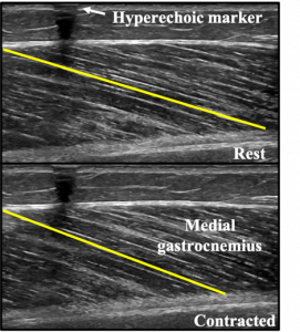
Left: Ultrasound images of the medial gastrocnemius muscle belly of a person with Parkinson’s disease at rest and during a contraction at 50% of their maximum strength. Measurement of the muscle is shown in yellow in the diagram, with the fiber shortening from the resting to contracted state during the contraction. The hyperechoic marker visible on the top of the image is used to ensure that the ultrasound probe does not move during the contraction.
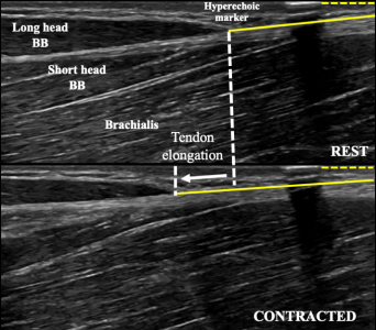
Right:Ultrasound of the muscle-tendon junction between the biceps brachii long head and the distal biceps tendon in a young male at rest and during a contraction at 25% of their maximum strength. Measurement of the distance from the muscle-tendon junction to the edge of the ultrasound screen in the resting and contracted state is shown in yellow and the difference between these two lengths is the measurement of tendon elongation.
Ultrasound of the muscle-tendon junction between the medial gastrocnemius and Achilles tendon in a person with Parkinson’s disease at rest and during a contraction at 50% of their maximum strength.
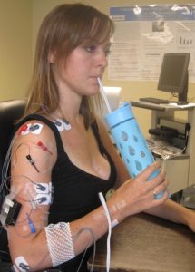
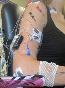
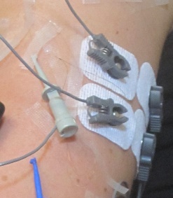
Electromyography (EMG)
EMG is a common tool through which researchers are able to visualize the electrical activity of the motor neurons (and consequentially, the muscles), in this case taken via a surface electrode placed non-invasively onto the skin.